Rat TNF-α ELISA kit

Promotion
Code: CSB-E11987r
Size:
96T,5×96T,10×96T
96T,5×96T,10×96T
Pls inquire
Quantity:
Sample Types: serum, plasma, cell culture supernates, tissue homogenates
Detect Range: 6.25 pg/mL-400 pg/mL
Sensitivity: 1.56 pg/mL
Trial Size:
24T ELISA Kit Trial Size (Only USD$150/ kit)
* Sample kit cost can be deducted as a $30 credit for each 96-assay kit of the same analyte and brand you subsequently purchase within six months until depleted. More details >>
Interested in a trial size? Please leave a message below.

For inquiries on large quantities or another requirements
Send an Inquiry
Start an on-line Chat
Online ordering is currently available for U.S. customers only. For orders outside the U.S., please kindly submit an inquiry or start a chat with us.
Promotion

Product Details

Alternative Names
Tnf ELISA Kit; Tnfa ELISA Kit; Tnfsf2 ELISA Kit; Tumor necrosis factor ELISA Kit; Cachectin ELISA Kit; TNF-alpha ELISA Kit; Tumor necrosis factor ligand superfamily member 2 ELISA Kit; TNF-a) [Cleaved into: Tumor necrosis factor ELISA Kit; membrane form ELISA Kit; N-terminal fragment ELISA Kit; NTF); Intracellular domain 1 ELISA Kit; ICD1); Intracellular domain 2 ELISA Kit; ICD2); C-domain 1; C-domain 2; Tumor necrosis factor ELISA Kit; soluble form] ELISA Kit
Abbreviation
TNF
Uniprot NO.
Species
Rattus norvegicus (Rat)
Sample Type
serum, plasma, cell culture supernates, tissue homogenates
Detection Range
6.25 pg/mL-400 pg/mL
Sensitivity
1.56 pg/mL
Assay Time
1-5h
Sample Loading Volume
50-100ul
Detection Wavelength
450 nm
Research Area
Immunology
Assay Principle
quantitative
The tag type will be determined during production process. If you have specified tag type, please tell us and we will develop the specified tag preferentially.
Measurement
Sandwich
Storage
Store at 2-8°C. Please refer to protocol.
Shelf Life
6 months
Lead Time
3-5 working days after you place the order, and it takes another 3-5 days for delivery via DHL or FedEx.
Note: All of our proteins are default shipped with normal blue ice packs, if you request to ship with dry ice, please communicate with us in advance and extra fees will be charged.
Precision
Intra-assay Precision (Precision within an assay): CV%<8%      
Three samples of known concentration were tested twenty times on one plate to assess.  
Inter-assay Precision (Precision between assays): CV%<10%      
Three samples of known concentration were tested in twenty assays to assess.    
             
Linearity
To assess the linearity of the assay, samples were spiked with high concentrations of rat TNF-α in various matrices and diluted with the Sample Diluent to produce samples with values within the dynamic range of the assay.
  Sample Serum(n=4)  
1:1 Average % 90  
Range % 87-96  
1:2 Average % 99  
Range % 95-104  
1:4 Average % 101  
Range % 97-105  
1:8 Average % 98  
Range % 93-107  
Recovery
The recovery of rat TNF-α spiked to levels throughout the range of the assay in various matrices was evaluated. Samples were diluted prior to assay as directed in the Sample Preparation section.
Sample Type Average % Recovery Range  
Serum (n=5) 85 80-92  
EDTA plasma (n=4) 99 95-104  
             
             
Typical Data
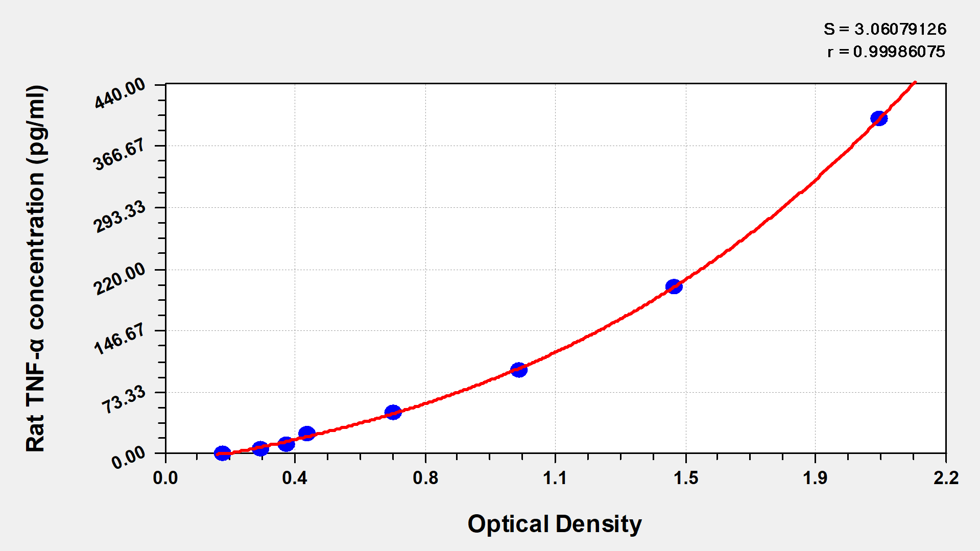
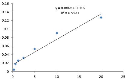
These standard curves are provided for demonstration only. A standard curve should be generated for each set of samples assayed.
pg/ml OD1 OD2 Average Corrected  
400 2.037 2.067 2.052 1.871  
200 1.445 1.489 1.467 1.286  
100 1.017 1.034 1.026 0.845  
50 0.655 0.677 0.666 0.485  
25 0.411 0.438 0.425 0.244  
12.5 0.351 0.378 0.365 0.184  
6.25 0.297 0.282 0.290 0.109  
0 0.183 0.179 0.181    
Protocols
/uploadfile/Ins/CSB-E11987r.pdf
Troubleshooting and FAQs
Description

Tumor necrosis factor (TNF) is a pro-inflammatory cytokine that regulates immune system function and inflammatory responses. First identified for its ability to kill tumor cells, TNF is mainly produced by activated macrophages and T cells, although many other cell types can secrete this cytokine. It acts as a key mediator in host defense against infections, autoimmune diseases, and cancer. TNF also contributes to the development of various inflammatory conditions. TNF produces its biological effects by binding to specific cell surface receptors, which triggers complex signaling cascades that regulate cell survival, proliferation, and apoptosis.

This rat TNF-α ELISA kit (CSB-E11987r) uses a quantitative sandwich measurement principle to detect tumor necrosis factor in Rattus norvegicus samples. The assay works with serum, plasma, cell culture supernates, and tissue homogenates with a detection range of 6.25 pg/mL to 400 pg/mL and sensitivity of 1.56 pg/mL. The protocol requires 50-100 μL sample volume and can be completed within 1-5 hours, with detection performed at 450 nm wavelength.

Application Examples

Note: The following application examples are drawn from a selection of publications citing this product. For additional applications, please refer to the full list of references in the "Citations" section.

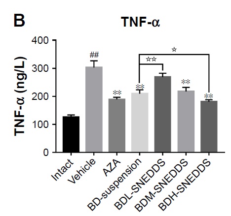
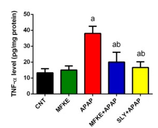
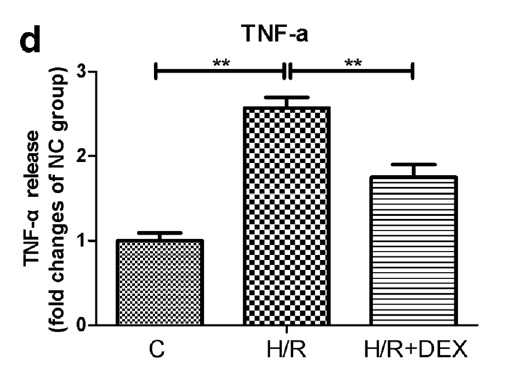
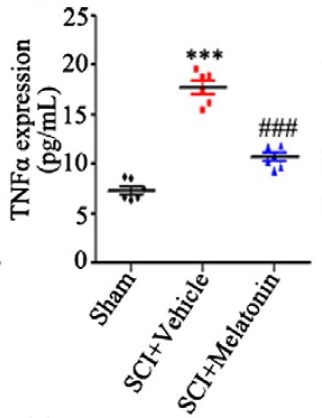
This rat TNF-α ELISA kit has been used across various research applications to quantify tumor necrosis factor-alpha levels in biological samples. The kit supports studies of inflammatory processes, metabolic disorders, and tissue-specific responses in rat models. Researchers have applied this assay to measure TNF-α concentrations in serum and tissue homogenates as part of comprehensive biomarker profiling studies.

Hepatic inflammation research - Quantification of TNF-α levels in liver tissue alongside other inflammatory mediators, apoptotic markers, and oncogenes to examine hepatic inflammatory responses
Metabolic disorder studies - Measurement of serum TNF-α concentrations in conjunction with insulin, adiponectin, and anti-inflammatory cytokines to evaluate metabolic dysfunction and inflammatory status
Antioxidant capacity research - Analysis of TNF-α levels alongside total antioxidant capacity and oxidative stress markers in serum samples from metabolic studies
Tissue-specific inflammation analysis - Evaluation of TNF-α content in adipose tissue and muscle homogenates as part of comprehensive inflammatory profiling in metabolic research
Multi-cytokine profiling - Simultaneous quantification of TNF-α with other pro-inflammatory mediators including prostaglandin E2 and interleukin-1β in inflammatory response studies
Biomarker correlation studies - Integration of TNF-α measurements with glycemic control markers, inflammatory mediators, and tissue-specific signaling molecules in comprehensive metabolic studies
Customer Reviews and Q&A

 Customer Reviews
Average Rating:
4.6 - 10 reviews

Submit a Review here

Sample species : a:1:{i:0;a:2:{i:0;s:3:"Rat";i:1;s:0:"";}}

Sample type: a:1:{i:0;a:2:{i:0;s:17:"Tissue homogenate";i:1;s:10:"PVN tissue";}}

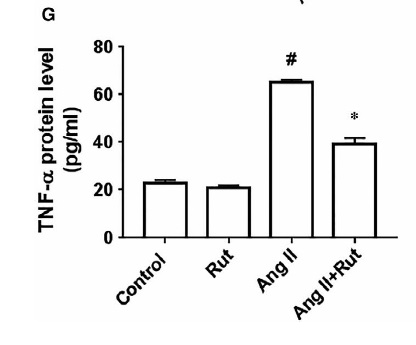
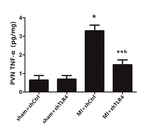
Review: The cytokine levels of IL-1β(F) and TNF-α(G) from the PVN tissue as measured by ELISA. n = 10–11 for each group. *P < 0.01 and **P < 0.05 versus sham groups; &P < 0.01 and #P < 0.05 versus MI + shCtrl group.

By Anonymous

Sample species : a:1:{i:0;a:2:{i:0;s:3:"Rat";i:1;s:0:"";}}

Sample type: a:1:{i:0;a:2:{i:0;s:24:"Cell culture supernatant";i:1;s:0:"";}}

Review: The cytokine levels of IL-1β(F) and TNF-α(G) from the PVN tissue as measured by ELISA. n = 10–11 for each group. *P < 0.01 and **P < 0.05 versus sham groups; &P < 0.01 and #P < 0.05 versus MI + shCtrl group.

By Anonymous

Sample species : a:1:{i:0;a:2:{i:0;s:3:"Rat";i:1;s:0:"";}}

Sample type: a:2:{i:0;a:2:{i:0;s:5:"Serum";i:1;s:0:"";}i:1;a:2:{i:0;s:17:"Tissue homogenate";i:1;s:11:"lung tissue";}}

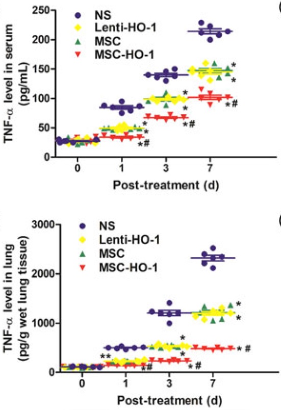
Review: The cytokine levels of IL-1β(F) and TNF-α(G) from the PVN tissue as measured by ELISA. n = 10–11 for each group. *P < 0.01 and **P < 0.05 versus sham groups; &P < 0.01 and #P < 0.05 versus MI + shCtrl group.

By Anonymous

Sample species : a:1:{i:0;a:2:{i:0;s:3:"Rat";i:1;s:0:"";}}

Sample type: a:1:{i:0;a:2:{i:0;s:5:"Serum";i:1;s:0:"";}}

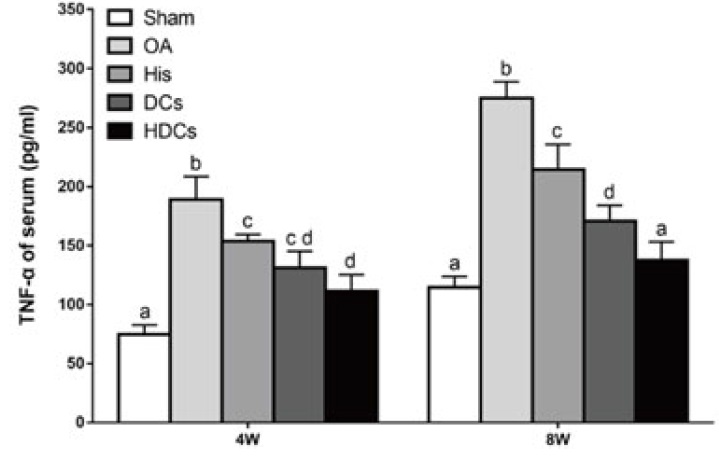
Review: The cytokine levels of IL-1β(F) and TNF-α(G) from the PVN tissue as measured by ELISA. n = 10–11 for each group. *P < 0.01 and **P < 0.05 versus sham groups; &P < 0.01 and #P < 0.05 versus MI + shCtrl group.

By Anonymous

Sample species : a:1:{i:0;a:2:{i:0;s:3:"Rat";i:1;s:0:"";}}

Sample type: a:1:{i:0;a:2:{i:0;s:17:"Tissue homogenate";i:1;s:18:"Colorectal tissues";}}

Review: The cytokine levels of IL-1β(F) and TNF-α(G) from the PVN tissue as measured by ELISA. n = 10–11 for each group. *P < 0.01 and **P < 0.05 versus sham groups; &P < 0.01 and #P < 0.05 versus MI + shCtrl group.

By Anonymous

Sample species : a:1:{i:0;a:2:{i:0;s:3:"Rat";i:1;s:0:"";}}

Sample type: a:1:{i:0;a:2:{i:0;s:17:"Tissue homogenate";i:1;s:16:"Liver homogenate";}}

Review: The cytokine levels of IL-1β(F) and TNF-α(G) from the PVN tissue as measured by ELISA. n = 10–11 for each group. *P < 0.01 and **P < 0.05 versus sham groups; &P < 0.01 and #P < 0.05 versus MI + shCtrl group.

By Anonymous

Sample species : a:1:{i:0;a:2:{i:0;s:3:"Rat";i:1;s:0:"";}}

Sample type: a:1:{i:0;a:2:{i:0;s:24:"Cell culture supernatant";i:1;s:0:"";}}

Review: The cytokine levels of IL-1β(F) and TNF-α(G) from the PVN tissue as measured by ELISA. n = 10–11 for each group. *P < 0.01 and **P < 0.05 versus sham groups; &P < 0.01 and #P < 0.05 versus MI + shCtrl group.

By Anonymous

Sample species : a:1:{i:0;a:2:{i:0;s:3:"Rat";i:1;s:0:"";}}

Sample type: a:1:{i:0;a:2:{i:0;s:17:"Tissue homogenate";i:1;s:18:"Spinal cord tissue";}}

Review: The cytokine levels of IL-1β(F) and TNF-α(G) from the PVN tissue as measured by ELISA. n = 10–11 for each group. *P < 0.01 and **P < 0.05 versus sham groups; &P < 0.01 and #P < 0.05 versus MI + shCtrl group.

By Anonymous

Sample species : a:1:{i:0;a:2:{i:0;s:3:"Rat";i:1;s:0:"";}}

Sample type: a:1:{i:0;a:2:{i:0;s:17:"Biological fluids";i:1;s:28:"Bronchoalveolar lavage fluid";}}

Review: The cytokine levels of IL-1β(F) and TNF-α(G) from the PVN tissue as measured by ELISA. n = 10–11 for each group. *P < 0.01 and **P < 0.05 versus sham groups; &P < 0.01 and #P < 0.05 versus MI + shCtrl group.

By Anonymous

Sample species : a:1:{i:0;a:2:{i:0;s:3:"Rat";i:1;s:0:"";}}

Sample type: a:2:{i:0;a:2:{i:0;s:5:"Serum";i:1;s:0:"";}i:1;a:2:{i:0;s:17:"Tissue homogenate";i:1;s:23:"heart tissue homogenate";}}

Review: The cytokine levels of IL-1β(F) and TNF-α(G) from the PVN tissue as measured by ELISA. n = 10–11 for each group. *P < 0.01 and **P < 0.05 versus sham groups; &P < 0.01 and #P < 0.05 versus MI + shCtrl group.

By Anonymous

 Q&A
Q:

Seeking for ELISA kits of TNF a .I am working on SD Rats and project related to High Fat Diet induced Metabolic complications.

A:
Thanks for your inquiry.
For the mentiond detection targets,we have the following kit:
#CSB-E11987r,Rat TNF-α ELISA kit
And we use kits to test ordinary rats.While there is not big problems to detect SD Rats.Pls take your actual test results as the real results
Q:

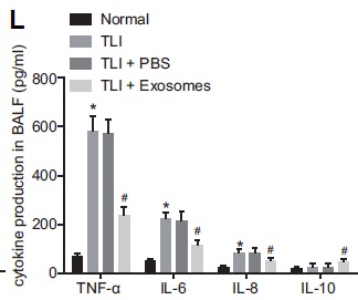
Rat TNF-α ELISA kit -CSB-E11987r this kit can be used for estimation in BALF...fluid
Kindly answer me.

A:
Thanks for your enquiry.
We usually use this kit to test serum, plasma, cell culture supernates, tissue homogenates.If you want to test bronchoalveolar lavage fluid,we suggest you do pre-testing first.If the content of your sample falls in the detection range,it can usually be detected out.
The manual will be provided to you by e-mail.
Pls let me know if you have any further questions. Thank you.
Q:

I want to know what the normal serum concentrations of TNFa in experimental rats are and the serum concentrations of TNFa in an induced freund adjuvant RA in experimental rats during 7 weeks. And I want to know the concentrations estimated in nano gramml. I want to ask about the weight loss of experimental animal rats also during an induced freund adjuvant arthritis RA during 7 weeks estimated by the gram.

A:

We used this CSB-E11987r Rat TNF-α ELISA kit to measure normal serum/plasma samples on rats. The model with heavy inflammation could even reach hundreds of pg/ml. The results varied with different conditions of the sample and different organism models. As to the mentioned induced model, we are not so sure about the sample condition. We suggest taking the actual measurement as your real result.
As to your question about weight loss, it usually depends on your own experiment aim or the choice of your target. If you just want to test TNF-α, then it is just ok to use the form of concentration of TNF-α.

Citations

Target Background

Function(From Uniprot)
Cytokine that binds to TNFRSF1A/TNFR1 and TNFRSF1B/TNFBR. It is mainly secreted by macrophages and can induce cell death of certain tumor cell lines. It is potent pyrogen causing fever by direct action or by stimulation of interleukin-1 secretion and is implicated in the induction of cachexia, Under certain conditions it can stimulate cell proliferation and induce cell differentiation. Induces insulin resistance in adipocytes via inhibition of insulin-induced IRS1 tyrosine phosphorylation and insulin-induced glucose uptake. Induces GKAP42 protein degradation in adipocytes which is partially responsible for TNF-induced insulin resistance. Plays a role in angiogenesis by inducing VEGF production synergistically with IL1B and IL6.; The TNF intracellular domain (ICD) form induces IL12 production in dendritic cells.
Subcellular Location
Cell membrane; Single-pass type II membrane protein.; [Tumor necrosis factor, membrane form]: Membrane; Single-pass type II membrane protein.; [Tumor necrosis factor, soluble form]: Secreted.; [C-domain 1]: Secreted.; [C-domain 2]: Secreted.
Protein Families
Tumor necrosis factor family
Database Links

  Email: support@cusabio.com
  Distributors Worldwide

CUSABIO guaranteed quality
icon of phone
Call us
301-363-4651 (Available 9 a.m. to 5 p.m. CST from Monday to Friday)
icon of address
Address
No. 269, Shendun 5th Road, Donghu Hi-Tech Development Area, Hubei Province, 430206, P.R.China
icon of social media
Join us with

Subscribe newsletter

Leave a message

* To protect against spam, please pass the CAPTCHA test below.
CAPTCHA verification
© 2007-2025 CUSABIO TECHNOLOGY LLC All rights reserved. 鄂ICP备15011166号-1
Select 0 Products