


Call us
301-363-4651 (Available 9 a.m. to 5 p.m. CST from Monday to Friday)

Digestive disorders are a group of various conditions that affect the structure and function of the digestive tract, including disorders of the esophagus, stomach, small intestine, large intestine, liver, gallbladder, pancreas, and other organs. These disorders can be caused by a variety of factors, including infections, autoimmune reactions, genetics, lifestyle and environmental factors. Symptoms of digestive disorders are varied, ranging from mild discomfort to severe pain, bleeding, or malnutrition. Common digestive disorders include gastritis, peptic ulcer, gastric cancer, hepatitis, cirrhosis, pancreatitis, and inflammatory bowel disease.
In recent years, significant progress has been made in the development of drugs for digestive diseases. For example, for the treatment of peptic ulcer, novel drugs such as the potassium ion-competitive acid blocker Vonorazan have been marketed, showing good efficacy and safety. In addition, for gastroesophageal reflux disease (GERD), the development of new acid-suppressing drugs is underway, aiming to provide better therapeutic efficacy and reduce side effects.
In the treatment of GI tumors, immunotherapy and targeted therapy technologies are rapidly evolving. For example, PD-1/PD-L1 antibodies and some targeted therapeutics (e.g., bevacizumab and cetuximab) have been approved for the treatment of advanced GI tumors.

Measured by its binding ability in a functional ELISA. Immobilized PD-1 at 2 μg/ml can bind Anti-PD-1 recombinant antibody, the EC50 of human PD-1 protein is 6.087-7.854 ng/ml.

Measured by its binding ability in a functional ELISA. Immobilized HER2 at 2 μg/ml can bind Trastuzumab, the EC50 is 2.179-2.825 ng/ml.

Human EGF protein captured on COOH chip can bind Human EGFR protein, his and Myc tag (CSB-MP007479HU) with an affinity constant of 11.9nM as detected by LSPR Assay.
-AC1.jpg)
Measured by its binding ability in a functional ELISA. Immobilized human CLDN18.2 (CSB-MP005498HU(A5)) at 5 μg/ml can bind anti-CLDN18.2 recombinant Monoclonal Antibody (CSB-RA005498A1HU), the the EC50 is 5.225-9.256 ng/ml.

Measured by its binding ability in a functional ELISA. Immobilized PD-L1 at 2 μg/ml can bind Anti- PD-L1 mouse monoclonal antibody (CSB-MA878942A1m, antigen from E.coli), the EC50 of human PD-L1 protein is 1.252-1.653 ng/mL

Measured by its binding ability in a functional ELISA. Immobilized CD152 at 2 μg/ml can bind Anti-CD152 rabbit monoclonal antibody (CSB-RA213310A0HU), the EC50 of human CD152 protein is 27.14-34.82 ng/ml.

Measured by its binding ability in a functional ELISA. Immobilized Human GLP1R at 2 μg/mL can bind Anti-GLP1R recombinant antibody (CSB-RA009514MA1HU), the EC50 is 54.54-94.23 ng/mL.

Measured by its binding ability in a functional ELISA. Immobilized TNFSF9 at 2 μg/ml can bind TNFRSF9(CSB-MP023984HU1), the EC50 is 2.671-3.702 ng/mL

CT26/Human GPC3 Stable Cell Line
Untransfected CT26 cells (green line) and transfected Human GPC3 CT26 Stable cells (red line) were stained with anti-GPC3 antibody (CSB-RA009705A1HU) (2µg/1*106 cells), washed and then followed by APC-conjugated anti-Human IgG Fc antibody and analyzed with flow cytometry.

HEK293T/Human CDH17 Stable Cell Line
Untransfected HEK293T cells (green line) and transfected Human CDH17 HEK293T stable cells (red line) were stained with anti-CDH17 antibody (CSB-RA613267MA1HU) (2µg/1*106 cells), washed and then followed by FITC-conjugated anti-Human IgG Fc antibody and analyzed with flow cytometry.

Untransfected HEK293T cells surface (green line) and transfected Human CDH17 HEK293T stable cells surface (red line) were stained with anti-CDH17 antibody (2µg/1*106 cells).
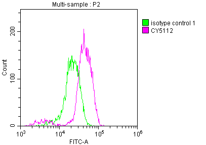
Overlay histogram showing Jurkat cells stained with CSB-RA159341A0HU (red line) at 1:50.

Overlay Peak curve showing MCF-7 cells stained with CSB-RA940669A0HU (red line) at 1:50.

Overlay Peak curve showing PC3 cells surface stained with CSB-RA215817A0HU (red line) at 1:100.
Tumour


